
The AMT Dental Assisting Radiography Certification Sample Question Set on this page is designed to familiarize you with the actual AMT DAR exam format and question types. These sample questions help you understand how questions are structured and what to expect on test day. While they provide a useful starting point, they represent only a limited preview of the real exam experience.
These sample questions are intended for evaluation and familiarization only. To understand exam style, pacing, and reasoning patterns more clearly, we recommend trying our online sample practice environment. If you are preparing for the AMT Dental Assisting Radiography (DAR) and want to assess your readiness more rigorously, structured, timed, scenario-based practice is recommended. This approach aligns with the cognitive demands and professional expectations typically associated with Dental Assistant, Dental Assisting Instructor, dental radiography support roles working in settings such as Dental offices, dental assisting education programs, military dental settings.
Try Sample Exam » | Access Full AMT DAR Practice Exam »
The demo introduces core concepts, while full-length premium simulations provide deeper, scenario-based coverage that more closely reflects the actual cognitive demands of the AMT Dental Assisting Radiography exam, particularly in areas such as dental radiography principles, imaging techniques and evaluation, radiation safety and infection control. You can use these sample questions as a starting point, then progress to the AMT DAR Certification Practice Exam for stronger readiness. Our premium simulations are designed to mirror real exam conditions, helping you refine reasoning, pacing, and decision-making before your official exam attempt.
AMT DAR Sample Questions:
01. Image/Graphic Stimulus Description: A four-panel diagram shows bitewing receptor placement options. Panel A shows the receptor centered over the crowns of maxillary and mandibular premolars with the occlusal plane level. Panel B shows the receptor placed too far anterior, cutting off the distal of the canine and missing distal premolars. Panel C shows the receptor tilted steeply so the occlusal plane is diagonal. Panel D shows the receptor placed vertically but positioned below the mandibular crowns only.
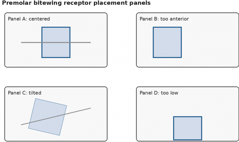
Which panel best demonstrates acceptable premolar bitewing receptor placement?
a) Panel A
b) Panel B
c) Panel C
d) Panel D
02. A dental assistant asks why the office adopted rectangular collimation for intraoral radiography even though it requires careful alignment.
Which explanation best supports the change?
a) Rectangular collimation eliminates all need for operator protection
b) Rectangular collimation reduces the area of tissue exposed when aligned properly
c) Rectangular collimation makes radiographs unnecessary for diagnosis
d) Rectangular collimation prevents every possible positioning error
03. A dental assistant is explaining why enamel, dentin, pulp, and restorations appear differently on a radiograph. The trainee asks why the final image has multiple shades rather than one uniform color.
Which explanation is most accurate?
a) Different tissues and materials absorb or transmit x-rays differently before the beam reaches the receptor
b) The receptor randomly assigns shades to structures after exposure
c) Dental x-rays become visible light only when they pass through enamel
d) All structures absorb the same amount of radiation, but processing changes their shape
04. A dental assistant is setting up a posterior periapical projection using the paralleling technique. The receptor is placed parallel to the long axis of the tooth, and a beam-alignment device is attached.
Which central-ray direction is most appropriate?
a) Parallel to the long axis of the tooth
b Directed steeply downward to intentionally foreshorten the roots
c) Perpendicular to both the tooth and the receptor
d) Directed only through the occlusal surfaces without regard to receptor position
05. Image/Graphic Stimulus Description: A labeled tube-head diagram shows the cathode filament on the left, the anode with a tungsten target on the right, the aluminum filter near the exit port, and the lead collimator closer to the position-indicating device. Electron arrows travel from the cathode filament to the tungsten target.
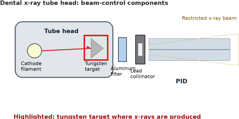
Based on the diagram, which component is the site where x-rays are produced after high-speed electrons strike it?
a) Tungsten target
b) Aluminum filter
c) Lead collimator
d) Position-indicating device
06. A dental assistant prepares an operatory for intraoral radiographs on a new patient. The x-ray control panel, receptor holder handles, and computer mouse are likely to be touched during the procedure.
Which preparation best reduces cross-contamination risk?
a) Leave surfaces uncovered because gloves alone prevent environmental contamination
b) Place appropriate barriers on surfaces that will be touched during exposure and replace them between patients
c) Disinfect only the dental chair and ignore radiography equipment surfaces
d) Use the same barriers for multiple patients if they do not appear visibly soiled
07. Image/Graphic Stimulus Description: A panoramic positioning diagram shows three patient positions relative to the focal trough. Position A places the anterior teeth centered within the focal trough. Position B places the anterior teeth too far forward. Position C places the anterior teeth too far backward.
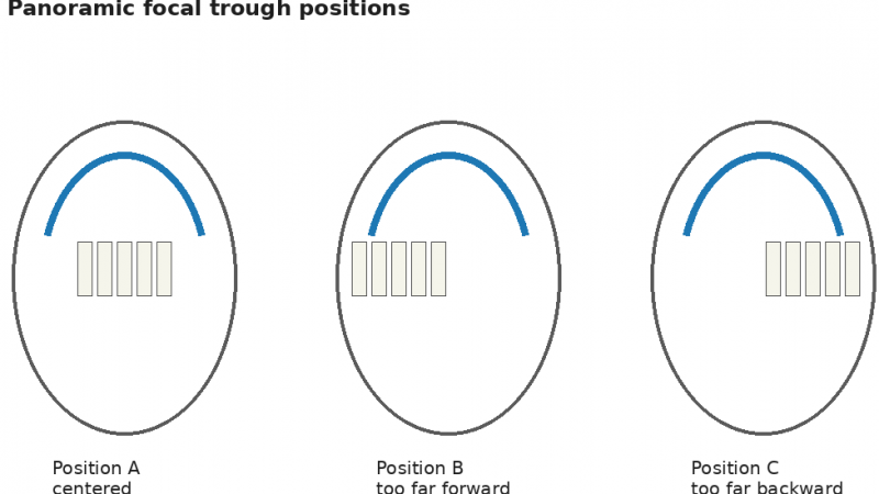
Which position is most likely to produce the sharpest anterior tooth image?
a) Position A
b) Position B
c) Position C
d) Any position produces equal sharpness if exposure time is increased
08. A dental office is transitioning from film to digital radiography. The dentist asks the assistant to explain one practical advantage of digital imaging for clinical workflow and quality assurance.
Which statement is most accurate?
a) Digital imaging eliminates all positioning errors during receptor placement
b) Digital sensors remove the need for infection-control barriers
c) Digital images can be viewed quickly and may be adjusted for brightness or contrast after acquisition
d) Digital imaging makes radiograph prescribing unnecessary because exposure is lower
09. A dentist requests an occlusal radiograph to evaluate a broad area of the maxillary arch. The assistant prepares to place the receptor intraorally so that a larger section of the arch can be recorded.
Which feature best distinguishes an occlusal projection from a standard periapical projection?
a) It records a larger area of the arch with the receptor positioned on the occlusal surfaces
b) It is used only to separate posterior interproximal contacts
c) It requires chemical fixation before exposure can occur
d) It eliminates the need to position the x-ray beam
10. A film radiograph is uniformly too dark. The assistant confirms that the film was processed for longer than the recommended developing time, while exposure settings were unchanged from prior diagnostic images.
Which processing factor most likely caused the dark image?
a) Inadequate fixation
b) Overdevelopment
c) Reversed film packet placement
d) Insufficient vertical angulation
Answers:
|
Question: 01 Answer: a |
Question: 02 Answer: a |
Question: 03 Answer: a |
Question: 04 Answer: c |
Question: 05 Answer: a |
|
Question: 06 Answer: b |
Question: 07 Answer: a |
Question: 08 Answer: c |
Question: 09 Answer: a |
Question: 10 Answer: b |
For full-length, timed, scenario-based practice aligned with the official exam framework - and to build pacing, consistency, and confidence - explore our Premium AMT DAR Certification Practice Exam.
Note: These sample questions are not official exam questions and are intended only for familiarization and study purposes. If you find any typos or data entry errors in these AMT Dental Assisting Radiography (DAR) sample questions, please let us know by emailing us at feedback@medicoexam.com