
CCI ACS Certification Sample Questions
The CCI Advanced Cardiac Sonographer Certification Sample Question Set on this page is designed to familiarize you with the actual CCI ACS exam format and question types. These sample questions help you understand how questions are structured and what to expect on test day. While they provide a useful starting point, they represent only a limited preview of the real exam experience.
These sample questions are intended for evaluation and familiarization only. To understand exam style, pacing, and reasoning patterns more clearly, we recommend trying our online sample practice environment. If you are preparing for the CCI Advanced Cardiac Sonographer (ACS) and want to assess your readiness more rigorously, structured, timed, scenario-based practice is recommended. This approach aligns with the cognitive demands and professional expectations typically associated with advanced cardiac sonographers, registered echocardiography sonographers, cardiovascular sonography professionals working in settings such as echocardiography laboratories, hospital cardiovascular imaging departments, cardiac diagnostic imaging settings.
Try Sample Exam » | Access Full CCI ACS Practice Exam »
The demo introduces core concepts, while full-length premium simulations provide deeper, scenario-based coverage that more closely reflects the actual cognitive demands of the CCI Advanced Cardiac Sonographer exam, particularly in areas such as advanced echocardiographic assessment, structural heart and interventional imaging, advanced and multimodality cardiac imaging. You can use these sample questions as a starting point, then progress to the CCI ACS Certification Practice Exam for stronger readiness. Our premium simulations are designed to mirror real exam conditions, helping you refine reasoning, pacing, and decision-making before your official exam attempt.
CCI ACS Sample Questions:
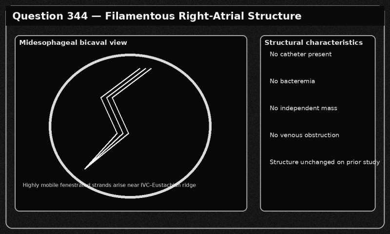
01. A highly mobile, filamentous, fenestrated structure arises from the inferior vena cava–Eustachian ridge region and moves freely within the right atrium. It has been unchanged on prior examinations. There is no catheter, bacteremia, venous obstruction, or independent mass.
Which interpretation is most appropriate?
a) Tricuspid-valve vegetation
b) Right-atrial myxoma
c) Chiari network, a benign embryologic right-atrial remnant/
d) Right-atrial thrombus in transit
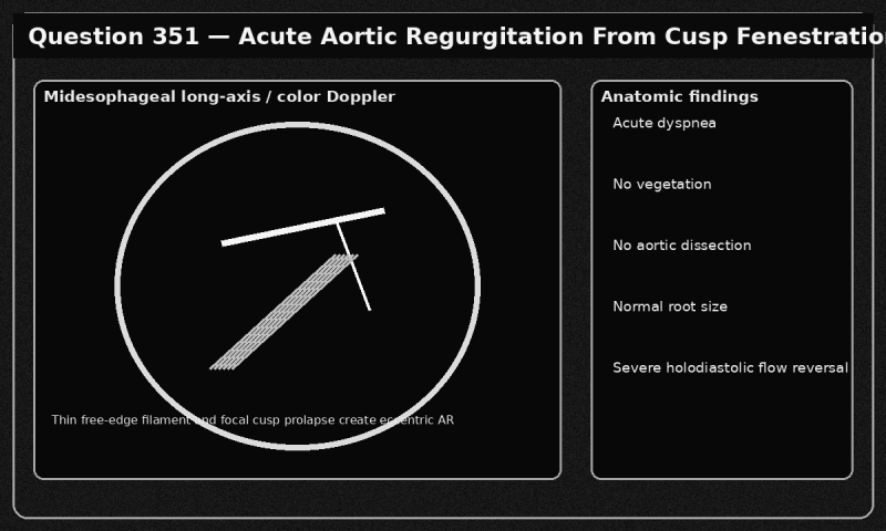
02. A patient develops acute severe aortic regurgitation. Transesophageal echocardiography shows a thin filament arising from the free edge of one aortic cusp, focal cusp prolapse, and an eccentric regurgitant jet. There is no vegetation, aortic dissection, or root dilation.
Which two conclusions are most appropriate? Select two correct responses.
a) Rupture of an aortic-cusp fenestration is the most likely mechanism/
b) The examination should define the affected cusp, coaptation defect, regurgitation severity, ventricular response, aortic root, and alternative acute causes, and the severe acute finding should be communicated urgently/
c) The eccentric jet proves chronic root-dilation–mediated regurgitation
d) The absence of a vegetation excludes a structural cusp lesion
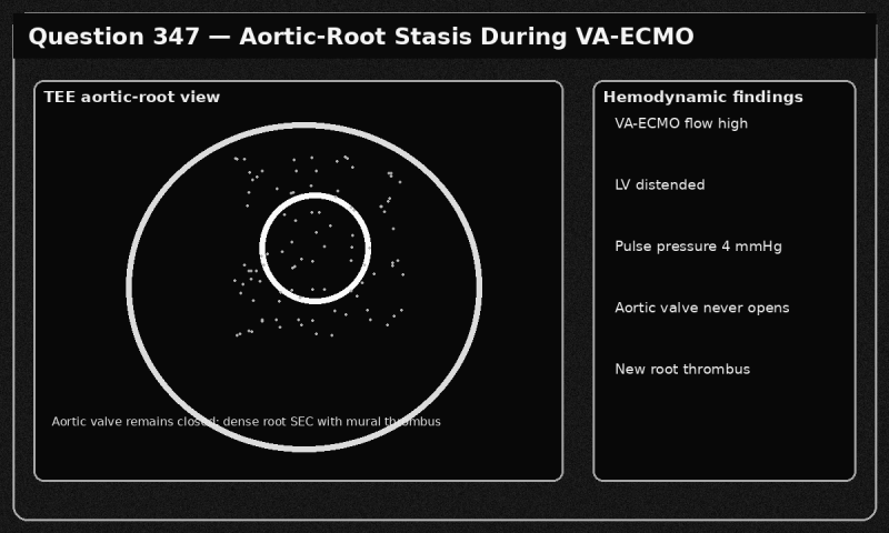
03. A patient receiving high-flow venoarterial extracorporeal membrane oxygenation has a distended left ventricle, pulse pressure of 4 mmHg, and an aortic valve that never opens. Transesophageal echocardiography shows dense spontaneous echo contrast and a new mural thrombus in the aortic root.
Which action is most appropriate?
a) Immediately communicate critical aortic-root stasis and thrombus and coordinate an urgent strategy to improve left-ventricular unloading and restore safe aortic-valve opening when feasible/
b) Ignore the thrombus because VA-ECMO prevents systemic embolization
c) Lower anticoagulation because spontaneous echo contrast indicates excessive blood thinning
d) Increase ECMO flow without reassessing left-ventricular loading
04. A patient with a transcatheter aortic valve has increasing exertional dyspnea. The prior study showed a mean gradient of 12 mmHg, Doppler velocity index of 0.52, and acceleration time of 75 ms. The current study shows a mean gradient of 30 mmHg, Doppler velocity index of 0.28, and acceleration time of 125 ms, but the preliminary report states, “No significant change.” Leaflet motion is poorly visualized.
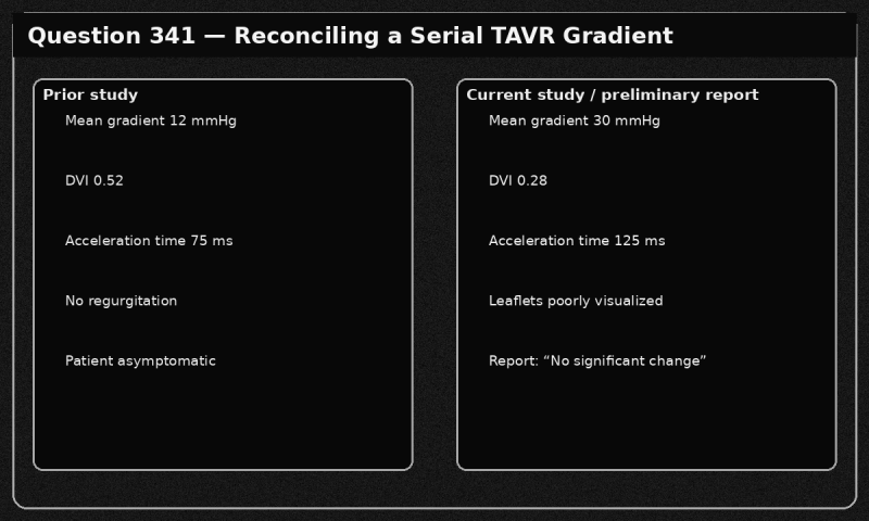
Which action is most appropriate?
a) Average the prior and current gradients and report the result as the present gradient
b) Delete the current Doppler measurements because the leaflets were not directly visualized
c) Keep the preliminary report unchanged because the current ejection fraction is not reduced
d) Reconcile the serial discrepancy, revise the preliminary interpretation, and promptly communicate possible new prosthetic obstruction requiring further evaluation/
05. A preliminary report calculates severe pulmonary hypertension from a dense 4.6-m/s apical continuous-wave signal. Review shows that the signal begins before the true tricuspid-regurgitation envelope and aligns with an eccentric mitral-regurgitation jet. The true TR envelope is incomplete and peaks near 2.8 m/s. Right-ventricular size and function, pulmonary-artery acceleration time, and inferior vena cava findings are normal.
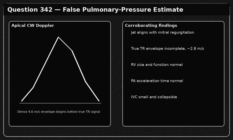
Which two actions are most appropriate? Select two correct responses.
a) Do not calculate right-ventricular systolic pressure from the contaminated 4.6-m/s signal/
b) Report severe pulmonary hypertension because the highest recorded velocity must always be used
c) Use the incomplete 2.8-m/s signal as an exact pressure measurement without qualification
d) Reacquire TR Doppler from multiple windows and integrate the best complete envelope with right-heart morphology, pulmonary-flow indices, and estimated right-atrial pressure/
06. After surgical left atrial appendage clipping, transesophageal echocardiography shows a residual patent stump measuring 15 mm in depth with color flow entering the pouch. The external clip is stable, no thrombus is present, and atrial fibrillation persists.
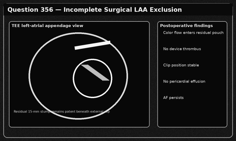
Which interpretation is most appropriate?
a) Complete appendage exclusion because the external clip is visible
b) Incomplete surgical left atrial appendage exclusion with a clinically relevant residual stump
c) Normal coronary-sinus flow
d) A new left-atrial dissection
07. An adult with right-heart volume loading has right-lung hypoplasia, cardiac dextroposition, a small right pulmonary artery, and a curved right pulmonary venous confluence descending below the diaphragm to the inferior vena cava. A secundum atrial septal defect is also present.
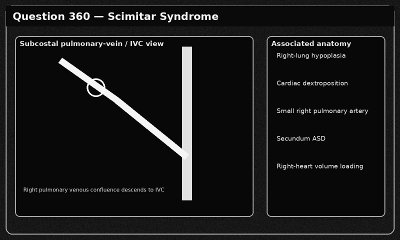
Which diagnosis is most consistent with the findings?
a) Total anomalous pulmonary venous return to a vertical vein
b) Superior sinus venosus atrial septal defect with right upper pulmonary vein drainage to the superior vena cava
c) Persistent left superior vena cava
d) Scimitar syndrome
08. Immediately after cardiopulmonary bypass, transesophageal echocardiography shows a new homogeneous mass projecting into the left atrium at the expected location of the left atrial appendage. The mass was absent before bypass, has no independent stalk, and the appendage is not visible externally.
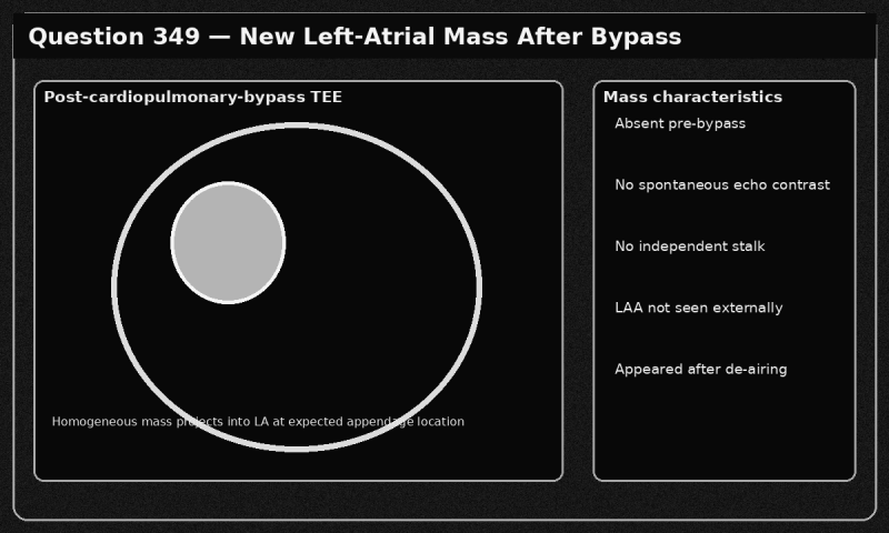
Which interpretation is most appropriate?
a) Acute left-atrial myxoma
b) Organized left-atrial thrombus
c) Inverted left atrial appendage
d) Left-atrial vegetation
09. In a patient with a small ascending aorta, Doppler echocardiography records an aortic-valve peak velocity of 4.3 m/s and mean gradient of 45 mmHg, while catheterization records a net mean gradient of 25 mmHg beyond the zone of recovered pressure. Doppler alignment is appropriate, stroke volume is normal, and no subvalvular obstruction is present.
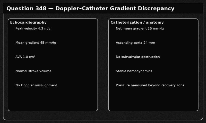
Which two conclusions are most appropriate? Select two correct responses.
a) The report should preserve the valid Doppler data while integrating valve area, flow state, aortic size, morphology, symptoms, and—when clinically relevant—energy-loss or invasive information
b) The catheter result proves that Doppler alignment was incorrect
c) The two gradients should be averaged to create the final severity grade
d) Pressure recovery can account for part of the Doppler–catheter gradient discrepancy, particularly when the ascending aorta is small/
10. The tricuspid leaflets are diffusely thickened with doming and restricted diastolic opening. At a heart rate of 70/min, the mean diastolic gradient is 7 mmHg. The right atrium is severely enlarged, the inferior vena cava is plethoric, and rheumatic mitral disease is present.
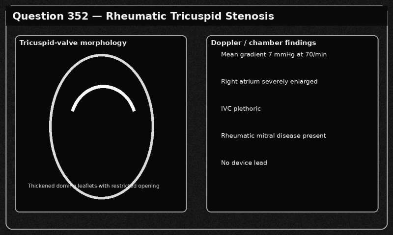
Which diagnosis is most consistent with the findings?
a) Normal tricuspid inflow variation
b) Hemodynamically important rheumatic tricuspid stenosis/
c) Carcinoid tricuspid regurgitation without stenosis
d) Functional tricuspid regurgitation
Answers:
|
Question: 01 Answer: c |
Question: 02 Answer: a, b |
Question: 03 Answer: a |
Question: 04 Answer: b |
Question: 05 Answer: a, d |
|
Question: 06 Answer: b |
Question: 07 Answer: d |
Question: 08 Answer: c |
Question: 09 Answer: a, d |
Question: 10 Answer: b |
For full-length, timed, scenario-based practice aligned with the official exam framework - and to build pacing, consistency, and confidence - explore our Premium CCI ACS Certification Practice Exam.
Note: These sample questions are not official exam questions and are intended only for familiarization and study purposes. If you find any typos or data entry errors in these CCI Advanced Cardiac Sonographer (ACS) sample questions, please let us know by emailing us at feedback@medicoexam.com


