
CCI RCS Certification Sample Questions
The CCI Registered Cardiac Sonographer Certification Sample Question Set on this page is designed to familiarize you with the actual CCI RCS exam format and question types. These sample questions help you understand how questions are structured and what to expect on test day. While they provide a useful starting point, they represent only a limited preview of the real exam experience.
These sample questions are intended for evaluation and familiarization only. To understand exam style, pacing, and reasoning patterns more clearly, we recommend trying our online sample practice environment. If you are preparing for the CCI Registered Cardiac Sonographer (RCS) and want to assess your readiness more rigorously, structured, timed, scenario-based practice is recommended. This approach aligns with the cognitive demands and professional expectations typically associated with Cardiac sonographers, cardiovascular technologists, diagnostic medical sonographers and related roles working in settings such as Hospitals, cardiovascular/echocardiography laboratories, outpatient diagnostic imaging centers.
Try Sample Exam » | Access Full CCI RCS Practice Exam »
The demo introduces core concepts, while full-length premium simulations provide deeper, scenario-based coverage that more closely reflects the actual cognitive demands of the CCI Registered Cardiac Sonographer exam, particularly in areas such as Echocardiographic imaging, cardiac/valvular/hemodynamic assessment, ultrasound physics and image optimization. You can use these sample questions as a starting point, then progress to the CCI RCS Certification Practice Exam for stronger readiness. Our premium simulations are designed to mirror real exam conditions, helping you refine reasoning, pacing, and decision-making before your official exam attempt.
CCI RCS Sample Questions:
01. A patient has suspected acute right ventricular infarction. Which two echocardiographic findings would support this diagnosis in the appropriate clinical setting? (Select two correct responses.)
a) New concentric left ventricular hypertrophy developing over several hours
b) Right ventricular dilation with elevated right-sided filling pressure
c) New regional or global right ventricular systolic dysfunction
d) Isolated severe left atrial enlargement with normal right ventricular function
02. A patient with acute inferior myocardial infarction is hypotensive with clear lungs. Echocardiography shows a dilated hypokinetic right ventricle, preserved left ventricular systolic function, and a plethoric inferior vena cava. Which diagnosis best explains the hemodynamic pattern?
a) Hypertrophic cardiomyopathy
b) Isolated acute left ventricular failure
c) Right ventricular infarction causing predominant right-sided pump failure
d) Severe chronic aortic regurgitation
03. A sonographer is differentiating left atrial appendage thrombus from normal anatomic structures on TEE. Which two features favor true thrombus? (Select two correct responses.)
a) Associated spontaneous echo contrast or markedly reduced appendage emptying velocity
b) A regular muscular ridge that follows the expected contour of normal pectinate muscle
c) A discrete mass that is visible in multiple imaging planes and remains distinct from the appendage wall and pectinate anatomy
d) A structure seen in only one plane that disappears completely with minor probe rotation
04. A sonographer suspects partial anomalous pulmonary venous return in a patient with unexplained right-sided volume overload. Which two findings would increase suspicion? (Select two correct responses.)
a) Failure to identify a normal pulmonary venous connection to the left atrium from a vein that appears to course toward a systemic vein
b) A small right ventricle with no evidence of increased pulmonary blood flow
c) A normal pulmonary venous connection pattern with all four veins clearly entering the left atrium
d) Right ventricular volume overload out of proportion to any identified intracardiac shunt
05. An adult with a large unrepaired ventricular septal defect has severe pulmonary hypertension. Color Doppler now shows bidirectional shunting with a dominant right-to-left component, and the VSD systolic velocity is much lower than on prior studies. Which hemodynamic process best explains these changes?
a) Systemic vascular resistance has fallen to zero while pulmonary resistance remains normal
b) The ventricular septal defect has become more restrictive and therefore produces lower velocity
c) The left ventricular systolic pressure has increased far above right ventricular pressure
d) Pulmonary vascular resistance has risen toward or above systemic resistance, producing Eisenmenger physiology
06. The accompanying apical image obtained after an inferior myocardial infarction shows a highly mobile structure attached to the mitral apparatus, loss of normal posterior leaflet support, and a broad eccentric systolic jet into the left atrium.
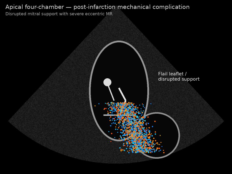
Which complication is most likely?
a) Stable chronic mitral valve prolapse
b) Normal papillary muscle motion
c) Papillary muscle rupture with flail mitral leaflet and acute severe mitral regurgitation
d) Mitral stenosis
07. The accompanying color Doppler image shows a large ventricular septal defect with low-velocity bidirectional flow. The right ventricle is hypertrophied and enlarged, and systolic septal flattening is present.
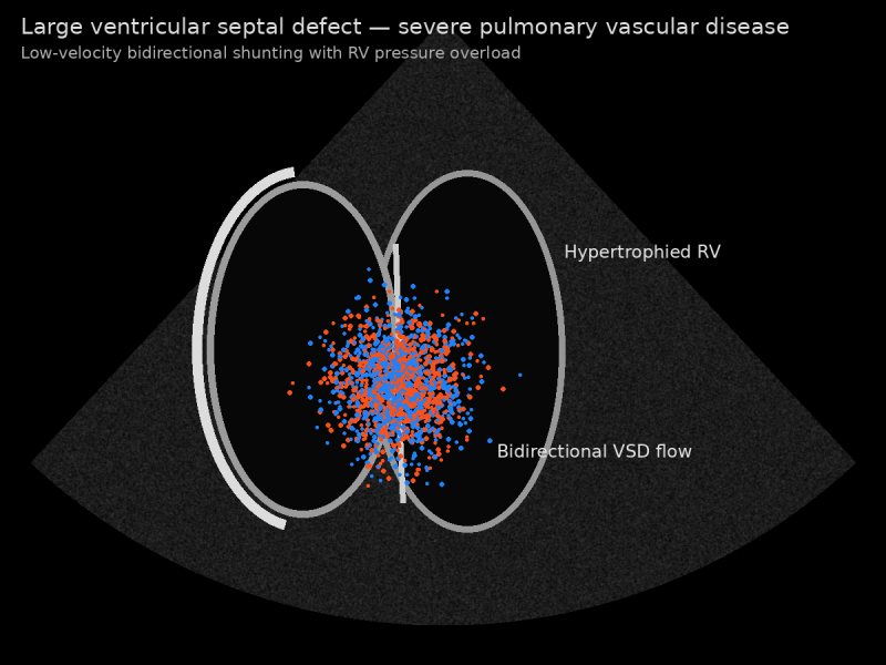
Which physiology is most consistent with these findings?
a) Normal physiologic interventricular flow
b) Eisenmenger physiology with near-systemic or systemic right ventricular pressure
c) A restrictive VSD with normal pulmonary artery pressure
d) Isolated left ventricular volume overload without pulmonary hypertension
08. Three days after an inferior myocardial infarction, a patient develops sudden pulmonary edema and a new loud systolic murmur. Echocardiography shows a hyperdynamic left ventricle and severe eccentric mitral regurgitation. Which mechanical complication should be strongly suspected?
a) Aortic dissection
b) Constrictive pericarditis
c) Papillary muscle rupture causing acute severe mitral regurgitation
d) Chronic secondary mitral regurgitation from gradual ventricular remodeling
09. A young adult with exertional symptoms has normal basal septal thickness but marked hypertrophy confined predominantly to the left ventricular apex. The apical cavity is small in systole and there is no resting LVOT gradient. Which cardiomyopathy phenotype is most likely?
a) Dilated cardiomyopathy
b) Apical hypertrophic cardiomyopathy
c) Arrhythmogenic right ventricular cardiomyopathy
d) Restrictive cardiomyopathy
10. A patient with exertional dyspnea has mitral E/A ratio of 1.4, septal e′ of 4 cm/s, lateral e′ of 6 cm/s, average E/e′ of 16, enlarged left atrial volume index, and tricuspid regurgitation velocity of 3.0 m/s. Which interpretation is most appropriate?
a) Normal diastolic function because the E/A ratio lies between 1 and 2
b) A pseudonormal transmitral pattern with evidence of elevated left ventricular filling pressure
c) Severe mitral stenosis because a high E/e′ ratio is specific for valvular obstruction
d) Isolated impaired relaxation with normal filling pressure
Answers:
|
Question: 01 Answer: b, c |
Question: 02 Answer: c |
Question: 03 Answer: a, c |
Question: 04 Answer: a, d |
Question: 05 Answer: d |
|
Question: 06 Answer: c |
Question: 07 Answer: b |
Question: 08 Answer: c |
Question: 09 Answer: b |
Question: 10 Answer: b |
For full-length, timed, scenario-based practice aligned with the official exam framework - and to build pacing, consistency, and confidence - explore our Premium CCI RCS Certification Practice Exam.
Note: These sample questions are not official exam questions and are intended only for familiarization and study purposes. If you find any typos or data entry errors in these CCI Registered Cardiac Sonographer (RCS) sample questions, please let us know by emailing us at feedback@medicoexam.com


