
CCI RPhS Certification Sample Questions

The CCI Registered Phlebology Sonographer Certification Sample Question Set on this page is designed to familiarize you with the actual CCI RPhS exam format and question types. These sample questions help you understand how questions are structured and what to expect on test day. While they provide a useful starting point, they represent only a limited preview of the real exam experience.
These sample questions are intended for evaluation and familiarization only. To understand exam style, pacing, and reasoning patterns more clearly, we recommend trying our online sample practice environment. If you are preparing for the CCI Registered Phlebology Sonographer (RPhS) and want to assess your readiness more rigorously, structured, timed, scenario-based practice is recommended. This approach aligns with the cognitive demands and professional expectations typically associated with venous/vascular sonographers, physicians and advanced practice clinicians, related vascular imaging professionals working in settings such as vascular ultrasound laboratories, phlebology/vein clinics, hospitals and outpatient diagnostic imaging settings.
Try Sample Exam » | Access Full CCI RPhS Practice Exam »
The demo introduces core concepts, while full-length premium simulations provide deeper, scenario-based coverage that more closely reflects the actual cognitive demands of the CCI Registered Phlebology Sonographer exam, particularly in areas such as venous diagnostic ultrasound, patient assessment and venous disease evaluation, venous treatment and procedure support. You can use these sample questions as a starting point, then progress to the CCI RPhS Certification Practice Exam for stronger readiness. Our premium simulations are designed to mirror real exam conditions, helping you refine reasoning, pacing, and decision-making before your official exam attempt.
CCI RPhS Sample Questions:
01. A lower-extremity venous Doppler tracing shows low-amplitude cardiac pulsatility in the common femoral vein on both sides, but the patient has no edema, dyspnea, or signs of right-sided heart failure. Which interpretation is most appropriate?
a) The finding proves bilateral acute DVT because normal venous flow is never pulsatile
b) Mild bilateral cardiac pulsatility can occur physiologically and should be interpreted with the overall clinical context
c) Any cardiac pulsatility proves severe tricuspid regurgitation
d) The waveform must be arterial contamination because venous waveforms cannot reflect cardiac activity
02. After completing a venous study, the sonographer steps away from the ultrasound workstation while the patient's name, date of birth, and images remain visible on the screen in an area accessible to other patients. Which action best protects patient information?
a) Leave the screen unchanged because no printed records are exposed
b) Turn the monitor toward the waiting area so the next patient can verify that the system is functioning
c) Lock or secure the workstation before leaving it unattended and follow the facility's privacy controls
d) Reduce monitor brightness but leave the patient record open
03. Immediately after ambulatory phlebectomy, the treating team applies a properly fitted compression dressing over the treated tributary sites. Which purpose best explains this routine step?
a) The dressing replaces all need for post-procedure observation or discharge instructions
b) Local compression can support hemostasis and help limit postoperative bruising and edema without intentionally occluding the deep venous system
c) Compression is applied only to create deep venous thrombosis so the superficial veins cannot refill
d) The dressing should permanently stop all arterial inflow to the limb
04. Refer to Image During evaluation of a suspected iliac venous lesion, intravascular ultrasound shows the catheter-centered cross-sectional lumen and reveals substantial focal compression that was underestimated on single-plane venography.

What is the main advantage of intravascular ultrasound in this setting?
a) It directly images the venous lumen and wall from within the vessel, improving assessment of lesion geometry and cross-sectional narrowing
b) It measures only superficial skin temperature around the groin
c) It is a noninvasive external ultrasound test performed without vascular access
d) It replaces all need for clinical history because intravascular images determine every cause of leg swelling
05. Refer to Image Immediately after sclerotherapy, a patient reports flashing lights and a transient scintillating visual pattern without focal weakness or speech disturbance. Vital signs are stable.

Which response is most appropriate?
a) Continue treatment because visual aura proves that the sclerosant has reached the target vein
b) Increase the sclerosant concentration so the visual symptoms resolve more quickly
c) Recognize the neurologic visual symptom, stop further injection, and promptly notify the treating clinician for assessment
d) Dim the room lights and discharge the patient immediately without clinical evaluation
06. Refer to Image In the deep lateral posterior calf, an artery is accompanied by paired veins coursing along the fibula and joining the tibial venous system proximally.
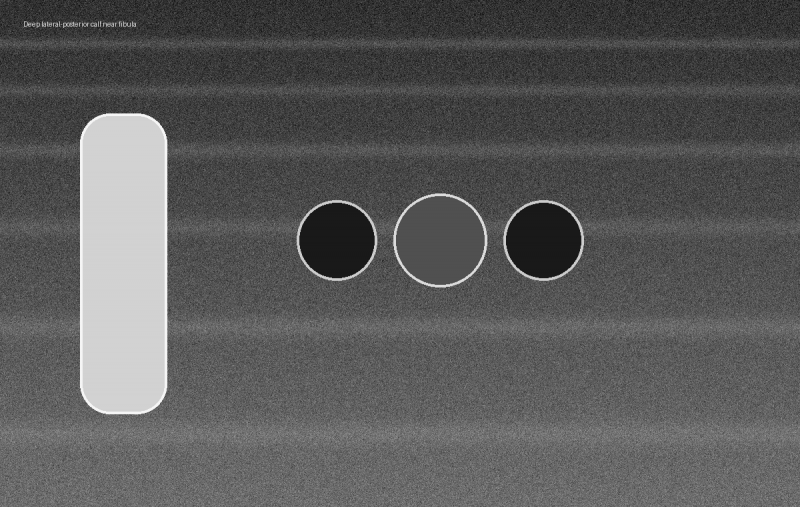
Which veins are being imaged?
a) Anterior saphenous veins
b) Cephalic veins
c) Great saphenous veins
d) Fibular (peroneal) veins
07. Refer to Image During thermal ablation, ultrasound shows a superficial target vein separated from the dermis by a newly created circumferential fluid layer after tumescent infiltration.
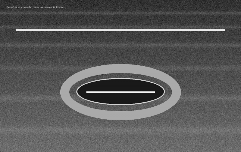
Which benefit of the image finding is most important?
a) The fluid layer increases physical and thermal separation between the treatment vein and the skin
b) The fluid layer proves that no ultrasound guidance is needed for the remainder of the procedure
c) The fluid intentionally obstructs the deep venous system to prevent blood return
d) The fluid converts thermal ablation into a chemical sclerotherapy procedure
08. Refer to Image During a reflux examination, color Doppler continues to display residual color in the vein for several frames after a brief provocative flow reversal has ended. The persistence control is set very high.
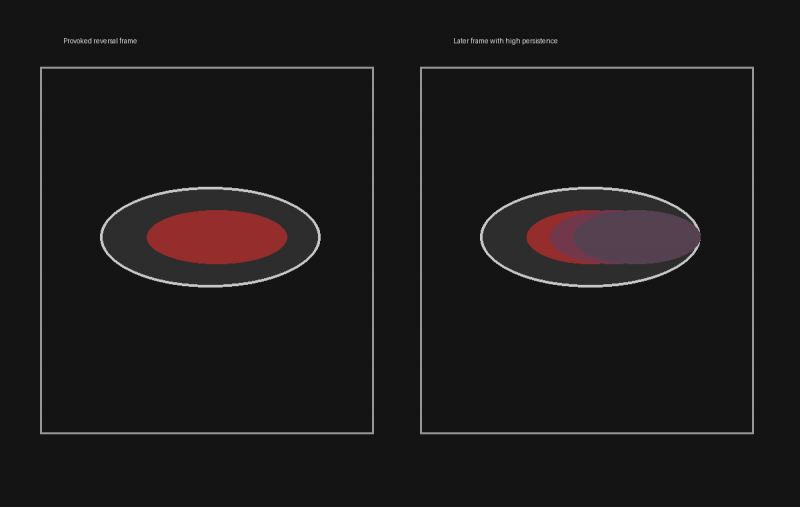
Which adjustment best reduces this temporal smearing?
a) Increase imaging depth because deeper imaging eliminates stored color information from prior frames
b) Increase persistence further so the reflux duration can be measured from a smoother display
c) Increase the wall filter because persistence artifacts are caused by low-frequency tissue motion
d) Decrease color persistence so fewer prior frames are averaged into the current display
09. A sonographer needs to measure Doppler velocity specifically within one focal portion of a venous stenosis rather than integrate velocities from the entire beam path. Which Doppler modality provides the needed range specificity?
a) Continuous-wave Doppler
b) Static grayscale cine review
c) Pulsed-wave Doppler
d) B-mode imaging alone
10. Before the first examination of the day, the sonographer notices that the ultrasound system's power cord has a split outer jacket with internal insulation visible near the plug. The system otherwise powers on normally. Which action is most appropriate?
a) Continue using the system because the internal conductors are not visibly exposed
b) Cover the damaged section with examination-table paper and continue scanning
c) Remove the system from clinical use and report the defect for qualified inspection or repair before further patient testing
d) Use the system only for noninvasive studies because electrical safety requirements apply only during invasive procedures
Answers:
|
Question: 01 Answer: b |
Question: 02 Answer: c |
Question: 03 Answer: b |
Question: 04 Answer: a |
Question: 05 Answer: c |
|
Question: 06 Answer: d |
Question: 07 Answer: a |
Question: 08 Answer: d |
Question: 09 Answer: c |
Question: 10 Answer: c |
For full-length, timed, scenario-based practice aligned with the official exam framework - and to build pacing, consistency, and confidence - explore our Premium CCI RPhS Certification Practice Exam.
Note: These sample questions are not official exam questions and are intended only for familiarization and study purposes. If you find any typos or data entry errors in these CCI Registered Phlebology Sonographer (RPhS) sample questions, please let us know by emailing us at feedback@medicoexam.com


