
CCI RVS Certification Sample Questions
The CCI Registered Vascular Specialist Certification Sample Question Set on this page is designed to familiarize you with the actual CCI RVS exam format and question types. These sample questions help you understand how questions are structured and what to expect on test day. While they provide a useful starting point, they represent only a limited preview of the real exam experience.
These sample questions are intended for evaluation and familiarization only. To understand exam style, pacing, and reasoning patterns more clearly, we recommend trying our online sample practice environment. If you are preparing for the CCI Registered Vascular Specialist (RVS) and want to assess your readiness more rigorously, structured, timed, scenario-based practice is recommended. This approach aligns with the cognitive demands and professional expectations typically associated with vascular sonographers, vascular ultrasound professionals, diagnostic medical sonography professionals and related roles working in settings such as hospitals and medical centers, vascular laboratories, diagnostic imaging facilities and related settings.
Try Sample Exam » | Access Full CCI RVS Practice Exam »
The demo introduces core concepts, while full-length premium simulations provide deeper, scenario-based coverage that more closely reflects the actual cognitive demands of the CCI Registered Vascular Specialist exam, particularly in areas such as vascular ultrasound examinations, ultrasound physics and vascular hemodynamics, patient care and safety. You can use these sample questions as a starting point, then progress to the CCI RVS Certification Practice Exam for stronger readiness. Our premium simulations are designed to mirror real exam conditions, helping you refine reasoning, pacing, and decision-making before your official exam attempt.
CCI RVS Sample Questions:
01. A basilic vein is patent and large enough for potential transposition, but it lies very deep beneath the skin along much of the upper arm. Why should depth be included in the mapping report?
a) Depth determines arterial stenosis severity
b) Depth is irrelevant once the vein is patent
c) Vein depth influences cannulation accessibility and the potential need for surgical transposition or superficialization
d) A deep basilic vein is always thrombosed
02. A long SFA segment is chronically occluded, with multiple collateral vessels reconstituting the popliteal artery and low-velocity monophasic distal flow. Which feature most strongly supports chronicity?
a) The presence of leg pain alone
b) An isolated elevated brachial pressure
c) Normal triphasic distal flow
d) Well-developed collateral reconstitution around the occluded segment
03. Before segmental pressure testing, a patient reports a severe latex allergy with a previous systemic reaction. The specialist is uncertain whether the pressure cuffs and tubing contain natural rubber latex. What is the safest next step?
a) Verify the equipment is latex-free or obtain approved latex-free alternatives before testing.
b) Proceed because vascular pressure cuffs do not contact mucous membranes.
c) Place a towel under the cuffs and use them without checking their material composition.
d) Premedicate the patient with an antihistamine and proceed with the available cuffs.
04. Refer to the image provided for this question. Longitudinal ICA imaging shows a heterogeneous plaque with an irregular crater-like surface defect extending into the plaque.
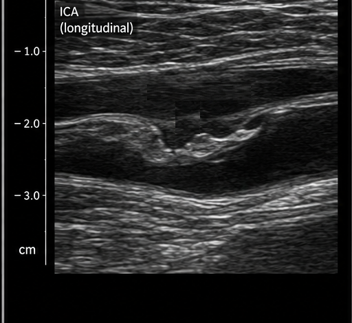
Which plaque feature is demonstrated?
a) Venous valve leaflet
b) Normal smooth intima-media complex
c) Complete ICA occlusion
d) Plaque ulceration or surface irregularity
05. Refer to the spectral Doppler image provided for this question. A long time window is displayed, compressing many cardiac cycles and making brief flow reversals difficult to evaluate.
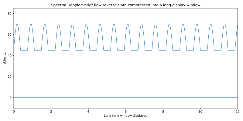
Which adjustment would best improve temporal detail of each cycle on the screen?
a) Increase spectral sweep speed so fewer cycles occupy the display width.
b) Increase color priority.
c) Increase B-mode dynamic range.
d) Decrease spectral sweep speed so more cycles fit on the screen.
06. Upper-extremity duplex shows an unusually high origin of the radial and ulnar arteries near the axilla rather than the usual antecubital bifurcation. Why is this finding important?
a) It means the brachial artery is occluded
b) It is an anatomic variant that should be documented because it changes expected vessel course and procedural planning
c) It is a venous anomaly only
d) It proves both forearm arteries are stenosed
07. Within hours of carotid stenting, the patient develops neurologic symptoms. Duplex shows echogenic material filling the stent and no internal color or spectral signal despite optimized settings. Which complication is most likely?
a) Subclavian steal
b) Normal post-stent flow
c) Mild nonocclusive restenosis
d) Acute carotid stent thrombosis
08. Refer to the image provided for this question.
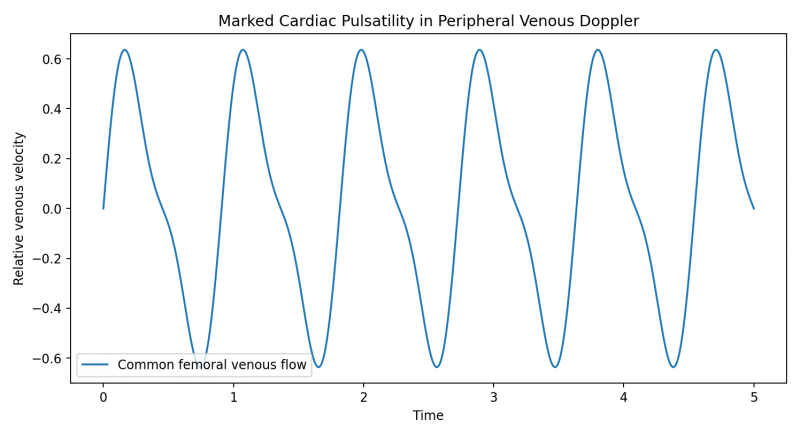
Both common femoral veins show strongly pulsatile spectral waveforms that closely reflect the cardiac cycle, and the veins remain compressible. Which systemic condition should be considered?
a) Acute bilateral femoral DVT
b) Isolated SFA stenosis
c) Elevated right-sided cardiac pressure or significant tricuspid regurgitation
d) Normal arterial collateralization
09. During a lower-extremity venous duplex examination for unilateral swelling, the specialist documents acute occlusive thrombus extending from the popliteal vein through the femoral vein. The patient is hemodynamically stable. What is the most appropriate next action after obtaining the necessary diagnostic documentation?
a) Complete the technical worksheet and allow the electronic report queue to notify the ordering clinician automatically.
b) Wait for the final signed report before any communication because preliminary findings should never be discussed.
c) Communicate the urgent finding promptly according to facility policy and document the date, time, and recipient of the communication.
d) Tell the patient that anticoagulation will be started and provide dosing instructions before contacting the medical team.
10. A color Doppler study of a rapidly pulsatile artery shows lingering color from prior frames, making brief changes in flow direction appear smeared over time. Which adjustment is most appropriate?
a) Increase color persistence.
b) Decrease color persistence.
c) Increase B-mode dynamic range.
d) Increase the wall filter to maximum.
Answers:
|
Question: 01 Answer: c |
Question: 02 Answer: d |
Question: 03 Answer: a |
Question: 04 Answer: d |
Question: 05 Answer: a |
|
Question: 06 Answer: b |
Question: 07 Answer: d |
Question: 08 Answer: c |
Question: 09 Answer: c |
Question: 10 Answer: b |
For full-length, timed, scenario-based practice aligned with the official exam framework - and to build pacing, consistency, and confidence - explore our Premium CCI RVS Certification Practice Exam.
Note: These sample questions are not official exam questions and are intended only for familiarization and study purposes. If you find any typos or data entry errors in these CCI Registered Vascular Specialist (RVS) sample questions, please let us know by emailing us at feedback@medicoexam.com


